Best Knee Arthroscopy in Rohini Delhi
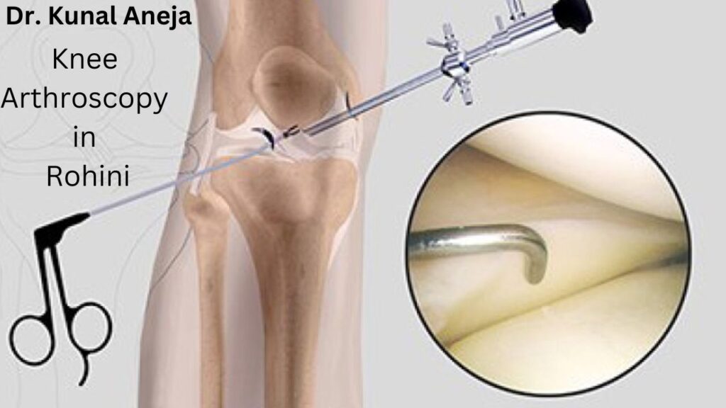
During the procedure of arthroscopy of the knee, a surgeon diagnoses and treats the joint problems with the knee. This type of surgery involves making a small incision and inserting a tiny camera called an arthroscopic camera into the knee. This camera allows them to view the inside of the knee on a screen, and is sometimes referred to as arthroscopic knee surgery.
It is well known that Naveda Healthcare Centre in Delhi have several top knee surgeons. Our hospital has a long history of treating patients who have developed knee-related conditions over the years. Our surgeons at our hospital are highly qualified and experienced, making it one of the best hospitals in India. Using the information that we have about the patient’s condition and medical history, we provide the best combination of treatment strategies to meet the patient’s needs.
How does it work?
A keyhole knee surgery is also known as an arthroscopic surgery and is used to treat severe osteoarthritis. The procedure usually results in problems walking, climbing stairs, and getting in and out of chairs.
The multidisciplinary teams at our hospital ensure our patients’ utmost care even after the surgery is completed, as we strive to address their knee pain successfully through the use of our multidisciplinary teams.
Recovering period
Within one or two weeks after surgery, most patients begin walking with a minimal limp, although full recovery takes four to six weeks.
Naveda Healthcare Centre As one of the best hospitals in Delhi, Dr. Kunal Aneja offers its patients specialized orthopedic surgery treatment in India, which includes comprehensive care and treatment. A systemic approach is used to treat a wide range of knee disorders and diseases. A team of experts in knee arthroscopy has been assembled at our practice, and we promise the best care to all our patients. As one of the first centers in Delhi to work round the clock, we treat our patients with minimal and affordable surgery costs, making us one of the best hospitals in India. During surgery, our patients have received excellent and desirable results.
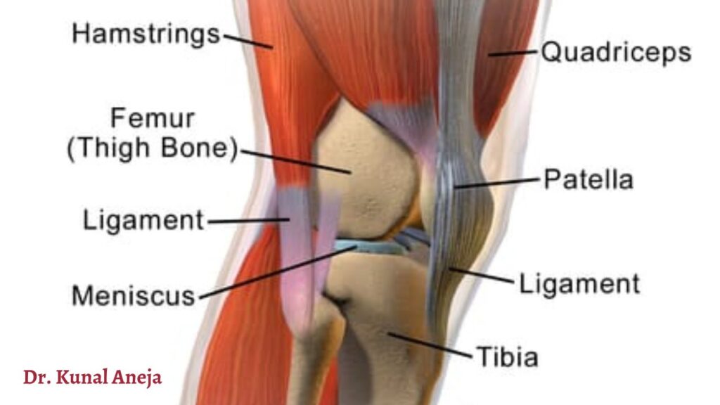
One of the most complex joints in the body is the knee joint, a simple hinge joint made up of the femur, tibia, and patella. The knee is a synovial joint, which means it is lined by synovium, which produces fluid that lubricates and nourishes the joint. Articular cartilage is the smooth surfaces at the ends of the femur and tibia. Articular cartilage is damaged, resulting in arthritis.
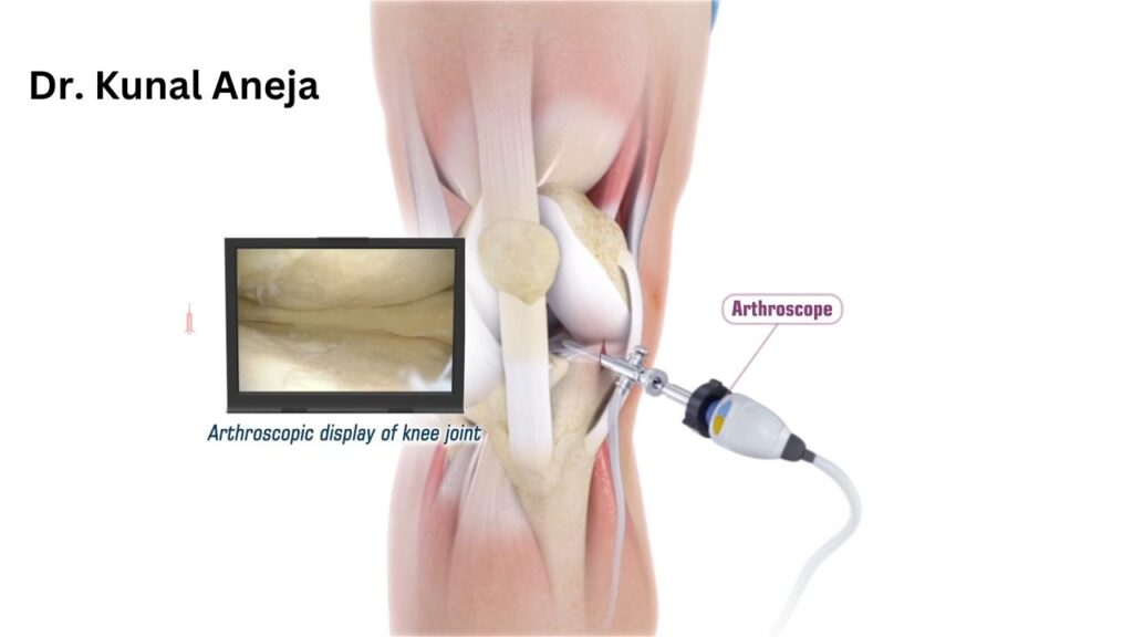
By definition, arthroscopy involves the insertion of an arthroscope into a joint, which derives from two Greek words, arthro-, meaning joint, and scopein, meaning to inspect.
The arthroscopy procedure is carried out in a hospital operating room.
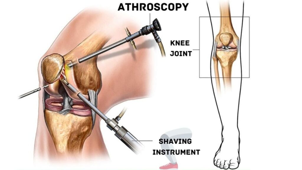
Arthroscopes are a piece of fiber optics used in arthroscopy surgery that has a tiny light source, a video camera, and a very small lens. Despite the fact that the surgical instruments used in one arthroscopy surgery are very small, they appear to be much larger when viewed through the arthroscope, which allows the viewer to see everything more clearly.
It is equipped with a television camera that displays an image of the joint on a television screen in order to be able to view the image of the joint on the television screen. Using the arthroscope, the surgeon can examine, for example, the cartilage and ligaments throughout the knee and under the kneecap with a television camera. Furthermore, a surgeon is also capable of repairing or correcting the problem, if it is necessary, as well as determining the extent or type of the injury.
As a result of thin incisions around the joint area (about 1/4 inch from the edge of the joint) the surgeon will be able to achieve the smoothest results possible. These incisions will produce very small scars that can be barely detected in most cases.
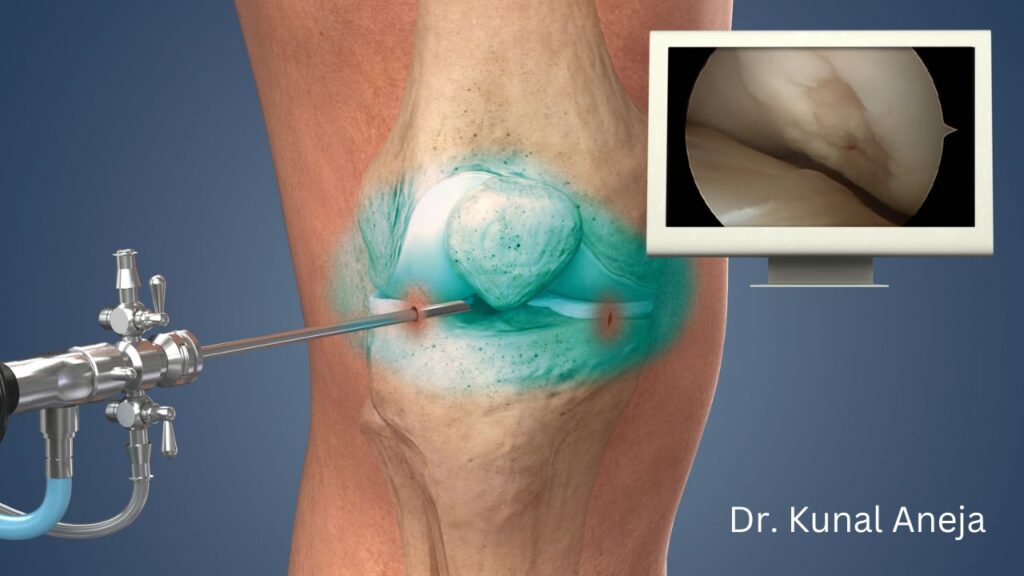
In order to observe the knee joint, surgeons insert an arthroscope into a portal. During the procedure, the surgeon also inserts an arthroscope, and then pumps a sterile solution into the knee joint. As a result of this sterile solution, the joint is expanded, which allows the surgeon to have a clearer view and more space to work in the joint. As part of the procedure, the amount of sterile solution that is injected into the joint can be controlled with a drainage needle.
By using the images created by the arthroscope, the surgeon may be able to guide himself/her in looking for any pathology or abnormalities in the body. By using a large image on a television screen, the surgeon is able to visualize the joint and determine the extent of the injury, thereby determining if surgery is required based on the size of the image on the television screen.
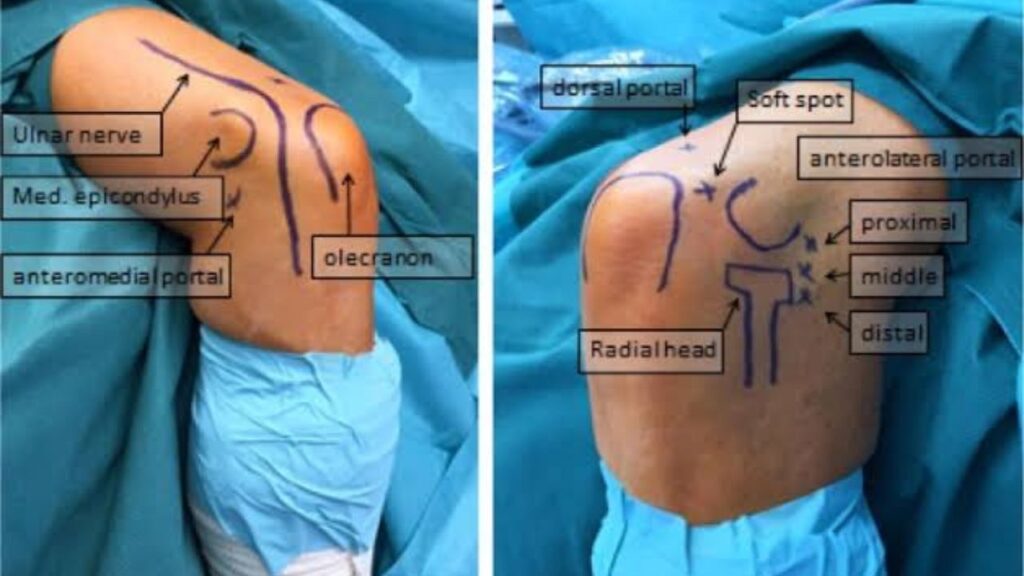
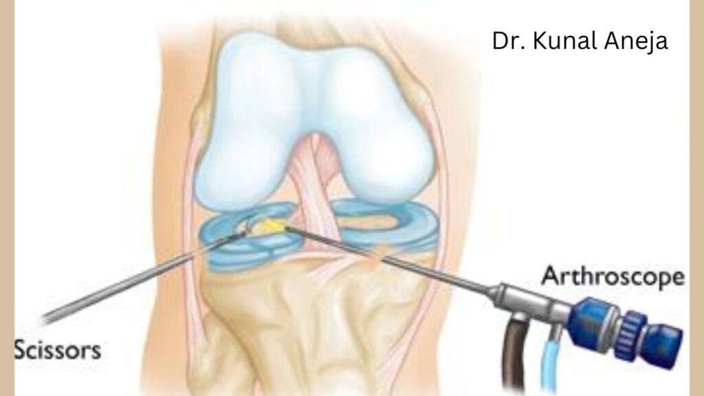
The other portal is used to insert surgical instruments so that if there is a problem with the joint, it will be possible for the surgeon to determine whether or not there is a problem with the joint by probing various parts of the joint. An excellent surgeon can utilize this portal (incision) to insert a variety of surgical instruments for treating any problems that may arise, and once the problem has been treated the portals (incisions) can be closed using suture or tape. It is important to understand that arthroscopy differs from traditional surgical methods where long incisions are made to open up the knees (arthrotomies) in that the muscles, ligaments, and tissues are not subjected to the same degree of trauma as they would be with an arthroscopy.
Why do we need arthroscopies?
A knee arthroscopy involves inserting a telescope into a small cut in your skin and looking inside the knee joint to diagnose problems. The instruments are then passed into the knee joint through several small cuts.
Is there any kind of anesthetic I can expect?
These types of surgeries require two types of anaesthesia:
You won’t feel anything during your operation if you are given a general anaesthetic – anaesthetic drugs that make you unconscious
A spinal anaesthetic is an injection in the lower back that numbs the lower half of your body. Sedation can be given accompanying the spinal anaesthesia.
A general anaesthetic is typically used for knee arthroscopies.
In the course of your consultation with your anesthetist, you will discuss the options for anesthesia – depending upon your health, age, and other medical conditions.
Types of treatments:
Treatment types include:
- Compared to traditional total knee replacements, laparoscopic knee replacements require a much smaller incision.
- The cruciate ligament can still be repaired after it has broken off from the bone if it still remains intact. It is attached to the bone again and held together with screws.
- Reconstruction of torn cruciate ligaments: Graft tissue is placed in the torn ligament in order to restore its function.
- An operation to repair the meniscus involves removing cartilage debris from the knee and stitching up tears.
- Knee cartilage is removed or smoothed out with chondroplasty.
- A partial knee replacement is carried out by removing only the damaged cartilage and ligaments in the knee, not the non-damaged areas.
What kind of arthroscopic knee replacement is appropriate for a patient depends on his or her condition.
Complications & Risks
As a surgical procedure, knee arthroscopy can sometimes result in some risks and complications, including:
- Thrombophlebitis
- Artery damage.
- Excessive bleeding
- Allergic reaction to anesthesia.
- Nerve damage.
- Numbness at the incision sites.
- Excessive pain in the calf and foot.
content by best healthcare marketing agency.

